
 Publish:2026-07-10
 Source:ReedBiotech
 Views:447
In the foundational chapter of our protein quantification series, we classified mainstream detection techniques into two core categories and illustrated the fundamental principles of non-specific total protein assays (UV absorbance, BCA assay, and Coomassie Brilliant Blue method) and conventional single-analyte specific detection approaches (ELISA, chemiluminescence assay, and TR-FRET), which sufficiently accommodate most routine experimental demands.
Nevertheless, practical biomedical research frequently encounters intractable technical limitations inherent to conventional methodologies, including the simultaneous quantification of over ten protein biomarkers per assay, the identification of ultra-low-abundance proteins beyond the detection limit of standard ELISA, and the synchronous measurement of multiple indicators with distinct dynamic ranges and sensitivity characteristics.
I. Multiplex Detection: Multi-Protein Analysis in a Single Assay – Efficient, Sample-Saving and Time-Efficient
Core advantages: This high-throughput multiplex detection platform enables simultaneous quantification of up to 500 target proteins in a single assay. It drastically reduces sample consumption, reagent dosage and overall experimental duration, effectively resolving common research bottlenecks including low detection efficiency and insufficient sample volume caused by repeated independent testing. It serves as an indispensable core tool for high-throughput biomedical research and large-scale clinical biomarker screening.
1. Luminex Assay (Liquid Phase Chip Technology)
Core principle: Color-coded microspheres are conjugated with target-specific antibodies. After sample incubation, target proteins bind to the corresponding microspheres, followed by the addition of fluorophore-conjugated detection antibodies to form immune complexes. The analyzer employs a dual-laser detection system: microsphere codes identify protein analytes, while fluorescence intensity reflects protein concentrations, enabling simultaneous quantification of multiple biomarkers from one single sample.
Key Experimental Workflow:Microsphere-sample incubation → addition of biotinylated detection antibodies → addition of streptavidin-phycoerythrin → instrumental detection. The assay requires less than 50 μL of sample and is fully compatible with 384-well plates.
Key advantages: Ultra-high throughput with over 500 detectable analytes per panel, high flexibility for customized biomarker panels, minimal sample consumption, and broad compatibility with complex biofluids such as serum and plasma.
Application scenarios: Simultaneous screening of multiple biomarkers (e.g., combined detection of inflammatory cytokines and tumor markers), high-throughput academic research, and combinatorial multi-index clinical analysis..
Limitations: The dedicated detection instrument carries a high upfront cost, associated reagents are expensive, and the assay must be operated by trained specialists.
2. Proximity Extension Assay (PEA)
Core principle: The Proximity Extension Assay (PEA) is an optimized upgraded technology derived from the Proximity Ligation Assay (PLA). Antibodies conjugated with specific oligonucleotides bind to target proteins, bringing paired complementary oligonucleotides into close proximity to achieve hybridization. DNA polymerase mediates the synthesis of double-stranded DNA templates, and target protein quantification is completed via microfluidic qPCR or NGS sequencing platforms.
Key Experimental Workflow: Incubation of oligonucleotide-conjugated antibodies with samples → Specific ligation reaction → PCR amplification → Instrument detection and quantitative analysis. The assay is applicable to multiple detection targets including human secreted proteins and organ-specific proteins.
Key advantages: Ultra-low detection limit at the fg/mL level, robust multiplex detection capability, superior adaptability to complex biological matrices such as plasma and cerebrospinal fluid, high detection specificity, and minimal sample volume requirement.
Application scenarios: Precision medicine research, early biomarker screening and identification, combined detection of inflammation-associated proteins, and preclinical drug target validation.
Limitations: Reliance on proprietary technical platforms with high reagent costs; operators are required to possess proficient qPCR experimental and analytical capabilities.
II. Ultra-Sensitive & Specialized Detection Technologies: Breaking Through Conventional Technical Limitations for Low-Abundance Protein Analysis
Technical advantages: These technologies exhibit significantly higher sensitivity than conventional detection methods, effectively overcoming technical bottlenecks in the detection of low-abundance proteins, single-cell sample analysis, and wash-free detection. They serve as essential technical supports for cutting-edge life science research and early clinical disease diagnosis.
1. Digital ELISA (Simoa, Single Molecule Array) – Gold Standard for Ultra-Sensitive Detection
Core principle: Based on the classic sandwich ELISA principle, the reaction system is diluted and distributed into millions of micro-wells, with each micro-well accommodating no more than a single protein molecule. Enzymatic substrate reactions generate specific fluorescent signals, which are captured by high-resolution fluorescence microscopy. According to the Poisson distribution theory, protein concentration is accurately calculated by quantifying the ratio of fluorescent positive micro-wells to the total number of bead-containing micro-wells.
Key Experimental Workflow: Incubation of antibody-coupled magnetic beads with samples → Addition of enzyme-labeled detection antibodies → Substrate reaction → Transfer to specialized detection discs → Automated CCD signal acquisition and analysis with full-process unmanned operation.
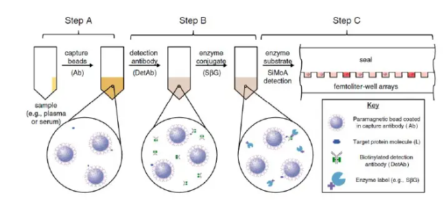
Key advantages: Ultra-high detection sensitivity reaching the fg/mL level, an ultra-broad dynamic range covering 4 orders of magnitude, simultaneous quantitative detection of more than 10 target molecules, and full-process automated operation.
Application scenarios: Detection of low-abundance proteins and trace biomarkers in cerebrospinal fluid and serum, early cancer screening, and quantitative detection of characteristic proteins associated with neurodegenerative diseases such as Alzheimer’s disease.
Limitations: Dependent on exclusive customized instruments and high-cost reagents; the technology is only applicable to experimental scenarios with ultra-high sensitivity detection requirements.
2. ELISpot (Enzyme-Linked Immunospot Assay) – Single-Cell Level Quantification
Core principle: ELISpot integrates cell culture technology with enzyme-linked immunosorbent assay. Target protein-secreting cells are seeded on antibody-coated microporous membranes, and secreted target proteins are captured in situ. Specific color reaction forms independent spots, where a single spot corresponds to a single protein-secreting cell. The frequency of secretory cells is quantitatively analyzed via spot counting.
Key Experimental Workflow: Microporous membrane coating with capture antibodies → Cell seeding and static culture → Addition of biotinylated detection antibodies → Addition of enzyme conjugates → Chromogenic reaction → Microscopic spot counting and analysis. Pre-experimental cell static treatment is required to effectively reduce false-positive signals.
Key advantages: Single-cell-level detection resolution, capable of functional evaluation of cellular secretory activity, with excellent detection sensitivity.
Application scenarios: Vaccine development and cellular immune response evaluation, tumor immune microenvironment monitoring, and auxiliary diagnosis of immunodeficiency diseases.
Limitations: The experimental procedure is cumbersome and requires stable cell culture conditions, relying on high-specificity antibodies; the technology is only applicable to the detection of secreted proteins.
3. PLA (Proximity Ligation Assay) – Exclusive Tool for Protein-Protein Interaction Analysis
Core principle: PLA technology innovatively combines specific antibody recognition with nucleic acid signal amplification. When the spacing between two target proteins is less than 30–40 nm, paired oligonucleotide-conjugated antibodies approach each other, enabling oligonucleotide ligation to form circular DNA templates. Rolling circle amplification (RCA) generates massive repeated nucleic acid sequences, and fluorescent staining produces distinct fluorescent puncta. The number of fluorescent spots corresponds to the abundance of target proteins or protein-protein interactions, realizing in-situ qualitative and quantitative analysis.
Key Experimental Workflow: Sample fixation and pretreatment → PLA probe incubation and specific binding → Enzymatic ligation to form circular DNA → Rolling circle amplification → Fluorescent staining → Microscopic imaging and quantitative analysis. The technology is compatible with cell slides and tissue section samples.
Key advantages: Ultra-high detection specificity and single-molecule-level sensitivity; it enables in-situ detection and quantitative analysis of protein interactions and post-translational modifications while preserving the original morphological structure of cells and tissues, filling the technical gap in precise quantification of protein-protein interactions.
Application scenarios:This technology is applicable to protein interaction verification, signal transduction pathway research, detection of various protein modifications, and quantitative determination of intracellular protein localization. Currently, based on this technology principle, our company has independently developed the protein quantitative immunoassay kit VeriQuant™.https://reedbiotech.com/view/304.html
Limitations: Complicated experimental operations, high reagent costs, and stringent requirements for antibody specificity; result interpretation requires solid professional biological knowledge and proficient fluorescence microscopy operation skills.
4. Mass Spectrometry (MS) — The “Gold Standard” for Precise Protein Quantification
Core Principle: Proteins isolated from biological samples are enzymatically digested into peptide fragments and subjected to ionization. Subsequent analyte separation and detection are performed based on mass-to-charge (m/z) ratios, with target protein quantification achieved via the peak intensity of signature peptides. The adoption of stable isotope-labeled peptide internal standards enables absolute quantitative analysis. Independent of protein spatial conformations, this methodology effectively eliminates analytical errors caused by epitope masking, a common limitation of antibody-dependent assays.
Key Experimental Workflow: The standardized workflow consists of sequential steps: sample protein extraction, tryptic digestion, peptide separation and purification, mass spectrometric detection, and bioinformatic analysis involving signature peptide matching and peak intensity calculation. Two prevailing quantitative strategies are widely utilized in proteomic research: label-free quantification and tandem mass tag (TMT) labeling. Compared with label-free workflows, TMT-based quantification provides higher analytical precision and minimizes missing data values.
Core Advantages: Mass spectrometry delivers high-accuracy quantitative capability and supports simultaneous quantification of thousands of proteins in a single assay. As an antibody-free analytical platform, it enables the detection and quantification of uncharacterized and unknown proteins. As an indispensable core tool for proteomic analysis, it is universally acknowledged as the gold standard for rigorous and reliable quantification of complex biological samples.
Application Scenarios: It is widely applied in comprehensive proteomic profiling, quantification of unknown proteins, screening of differentially expressed proteins, rigorous validation of core experimental results, and large-scale protein spectrum analysis.
Limitations: The instrument is expensive (costing millions), with complex operation and extremely stringent requirements for sample processing (including impurity removal and thorough enzymatic hydrolysis). It has a long detection cycle, requires professional personnel for operation and data analysis, and is limited in the detection of low-molecular-weight proteins.
III. Advanced Chapter Summary – Quick Reference Guide
1. Multiplex detection: Employ the “Luminex platform” for efficient high-throughput profiling, and the “PEA methodology” for ultrasensitive quantification of trace analytes.
2. Ultra-sensitive detection: Employ the “Simoa digital ELISA” to achieve ultrahigh analytical sensitivity.
3. Specialized scenario detection: Employ ELISpot for single-cell resolution analysis, PLA for protein-protein interaction profiling, and mass spectrometry for unbiased proteomic profiling.
The sophisticated analytical methodologies elaborated in this advanced chapter are capable of resolving 99% of intractable quantitative research bottlenecks. Investigators frequently raise the question of how to rapidly screen the optimal analytical workflow compatible with their experimental designs. Readers are encouraged to follow our forthcoming practical chapter, which will deliver definitive guidelines encompassing rational assay selection and systematic troubleshooting strategies to mitigate experimental artifacts and expedite robust data generation.