
 Publish:2026-11-06
 Source:ReedBiotech
 Views:110
With the same kit and the same batch of samples, why does someone else get beautifully blue wells while your results seem a little off? The color may be too faint, and the background may not be clean enough. The overall result just doesn't look right. Many people put this down to “experimental magic.” But color development is not a matter of luck. It mainly depends on three factors: the efficiency of electron transfer in the HRP catalytic cycle, the extent to which TMB is oxidized, and the stop solution added at the end.
Today, we're not going to discuss the operating procedures. Instead, we'll break down the underlying principles. It's important not only to know what happens, but also why it happens.
01 Let's Get to the Bottom of Signal Amplification
The principle of enzyme labeling can be summed up in one sentence: an enzyme is covalently conjugated to an antibody to form an enzyme-labeled antibody complex. The enzyme then specifically catalyzes its substrate, producing a colored insoluble product or particles with a certain electron density. These products can be detected by the naked eye, a light microscope, a microplate reader, or an electron microscope—in this way, the immune reaction becomes “visible.”
In 1966, Nakane and Pierce used horseradish peroxidase (HRP) instead of a fluorescent dye to localize antigens in tissues, pioneering the enzyme-labeled antibody technique. In 1971, Engvall and Perlmann built on this work to develop solid-phase enzyme-linked ELISA. At almost the same time, Van Weemen and Schuurs established an enzyme immunoassay using HRP-labeled HCG. Enzyme immunoassay thus became the third generation of immunolabeling technology, following radioimmunoassay and immunofluorescence. As monoclonal antibody technology matured, direct, indirect, and double-antibody sandwich methods were developed.
However, not every enzyme that can produce a color reaction is suitable as a label. It must meet all five of the following requirements:
① Substrate-specific, with products that are easy to observe under a light or electron microscope;
② The final product must be stable and must not diffuse into the surroundings or degrade;
③ The enzyme itself must be readily available and stable in its properties;
④ Conjugation with an antibody or antigen must not impair the activity of either;
⑤ The sample being tested must not contain a homologous endogenous enzyme.
Very few enzymes meet all these requirements. Only three have emerged as widely used options: HRP (horseradish peroxidase), ALP (alkaline phosphatase), and β-GAL (β-galactosidase). HRP is easy to extract, stable in its properties, and loses little activity after conjugation, making it a widely used choice for ELISA, WB, and reverse dot blot hybridization.
Let's bring the focus back to ELISA itself. Taking the most commonly used double-antibody sandwich method as an example, only one step in the entire plate is actually responsible for “visualization”:
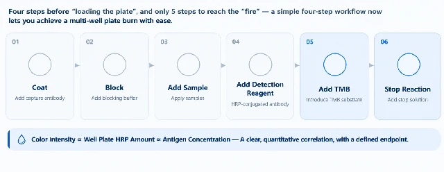
Figure 1. The six steps of a double-antibody sandwich ELISA. The first four steps determine how much enzyme is retained; the fifth step determines how strong the color becomes.
Pay attention to the order of cause and effect: color intensity is proportional to the amount of HRP remaining in each well, and the amount of residual HRP is proportional to the antigen concentration. If any of the first four steps is not performed properly, the fifth step will faithfully amplify the problem into “background” or “deviation.” This proportional relationship is the foundation of all quantitative results.
02 HRP: Why This Enzyme?
HRP is widely distributed throughout the plant kingdom, with the highest content found in horseradish (Armoracia rusticana), a member of the Brassicaceae family. It consists of a colorless enzyme protein and a brown iron-porphyrin group. Its Enzyme Commission number is EC 1.11.1.7. It is readily soluble in water (5% W/V), and its solution is transparent and reddish-brown.
Each HRP molecule contains one iron(III) protoporphyrin IX prosthetic group. The prosthetic group has a maximum absorbance at 403 nm, while the apoenzyme has a maximum absorbance at 275 nm. The ratio of these two absorbance values, OD403/OD275, is what is commonly known as the RZ value.
An RZ value above 3 indicates high quality, while purification is recommended when the RZ value is below 2.5. However, RZ only reflects the proportion of heme groups in the enzyme; it is not equivalent to actual purity and has no direct correlation with enzyme activity. Both activity and RZ should be considered when selecting an enzyme.
HRP has at least seven isoenzymes, with isoelectric points ranging from pH 3.0 to 9.0. Its optimum pH is usually around 5.0. In 1997, the crystal structure of HRP C was resolved at 2.15 Å [5]. It is a single-subunit glycoprotein containing 308 amino acids and eight carbohydrate chains, with a relative molecular mass of approximately 44 kDa. Its catalytic behavior, substrate preferences, and stability are all encoded in this structure.
What truly makes HRP indispensable is the catalytic cycle described below.
03 The Catalytic Cycle: A “Single-Electron” Relay
The active center of HRP is an iron protoporphyrin. Its catalytic process consists of three steps and passes through two high-oxidation states :
Step 1: Resting-state HRP (Fe³⁺) reacts with H₂O₂ and is oxidized to Compound I. The iron becomes Fe(IV)=O, while the porphyrin ring loses one electron to form a π-cation radical. This step is equivalent to storing the two oxidizing equivalents from H₂O₂ in the enzyme.
Step 2: The substrate enters and donates one electron, reducing Compound I to Compound II: Fe(IV)=O.
Step 3: The substrate donates another electron, and the enzyme returns to its resting state. The cycle is complete.
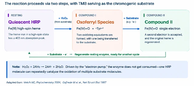
Figure 2. The three-step catalytic cycle of HRP.The substrate transfers electrons to the enzyme one at a time in two separate steps.This is the reason TMB can produce two different colors.
The net reaction is H₂O₂ + 2AH₂ → 2AH· + 2H₂O. Please note two details:
First, the substrate donates its two electrons separately. Therefore, the intermediate product must be a free radical—and the subsequent fate of this radical directly determines the color you observe.
Second, the enzyme is not consumed. A single HRP molecule can repeatedly initiate the cycle thousands of times. So-called “signal amplification” does not mean simply making the signal bigger; it means repeatedly “copying” a single binding event. This is why even a very small amount of enzyme-labeled antibody remaining in a well can ultimately produce a clearly measurable OD value.
04 TMB: A Substrate That Changes Color Twice
When TMB (3,3′,5,5′-tetramethylbenzidine) is first added to a well, it is colorless. Once HRP starts working, TMB is oxidized in two successive stages, and its color changes twice.
During the first oxidation, TMB loses one electron and becomes a radical cation. This species pairs with unoxidized TMB to form a diamine-diimine charge-transfer complex, which is deep blue and has a maximum absorbance at 652 nm. It remains in rapid equilibrium with the radical cation. Upon a second oxidation, diimine is formed. It is yellow, has a maximum absorbance at 450 nm, and is stable under acidic conditions.
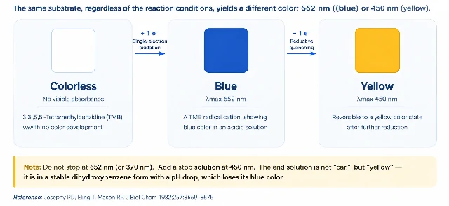
Figure 3. The same substrate produces different product structures at different oxidation levels, shifting the absorption peak from 652 nm to 450 nm.
A study by Josephy and colleagues, published in the Journal of Biological Chemistry in 1982, clearly explained this system: when H₂O₂ is insufficient, four forms—diamine, radical cation, charge-transfer complex, and diimine—exist in equilibrium. Adding an equimolar amount of H₂O₂ shifts the system entirely toward yellow diimine. This also explains two “rules” in ELISA:
Without stop solution → Read at 652 nm (or 370 nm)
The blue color continues to accumulate. This is a kinetic measurement with a short reading window, so the plate must be read quickly.
With stop solution → Read at 450 nm
The acid shifts the equilibrium toward stable diimine, changing the color from blue to yellow. The absorption shifts to a longer wavelength, the background is lower, and the plate can be read less urgently.
Therefore, the stop solution is not simply a “brake”; it is more like a “fixative.” It performs two functions at the same time: it completely inactivates the enzyme and stops further catalysis, while locking the product into a stable chemical form. Once you understand this, it becomes clear why the plate should be read as soon as possible after adding the stop solution. Although yellow diimine is stable, it does not remain valid indefinitely. Reading within 5 minutes is generally recommended.
05 Why Has TMB Become the Mainstream Chromogenic Substrate?
Before TMB, benzidine-based chromogens were widely used. They produced attractive colors, but were carcinogenic. In 1981, Bos and colleagues introduced TMB into enzyme immunoassays and clearly reported that it was a non-mutagenic chromogen with a negative Ames test. In sandwich EIAs for HCG and HBsAg, and in a competitive EIA for testosterone, the color yield of TMB also exceeded that of OPD (o-phenylenediamine), which was the best-performing substrate at the time .
TMB replaced benzidine and OPD because it combined three qualities: sensitivity, stability, and safety. The “blue” in this context is not a marketing term; it represents a genuine upgrade in chromogen safety and regulatory compliance.
06 Three Key Factors Affecting Color Development
Why can the same kit produce such different results in different users' hands? Because color development is influenced by three key factors at the same time.
① H₂O₂ concentration. It is a double-edged sword: too little, and color development is slow and the signal remains weak; too much, and non-enzymatic oxidation increases (TMB itself can also be slowly oxidized by H₂O₂), raising the overall background. Worse still, excess H₂O₂ can drive HRP toward Compound III, an enzymatically inactive state. This is why Substrate Solution A and Substrate Solution B must be prepared separately and mixed immediately before use. If they are mixed and left for two hours, part of the substrate will effectively have been “used up” in advance.
② pH. The optimum pH for HRP is approximately 5.0. Acidic stopping conditions stabilize diimine, but if the reaction system is too alkaline, the blue product will fade on its own. This is why the washing buffer formulation and stop solution concentration should not be changed casually.
③ Time and temperature. Color development is an enzyme-catalyzed reaction. As a general rule of thumb, the reaction rate approximately doubles for every 10°C increase in temperature. Variations in room temperature, placing the plate near an air-conditioning outlet, or stacking plates during incubation can all lead to differences between wells.
07 What Causes Common Color Development Problems?
When we connect the points discussed in the previous sections, the frustrating phenomena encountered during experiments become easier to explain:
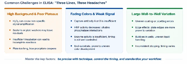
Figure 4. All abnormalities can be traced back by following three factors: “enzyme amount – substrate – time.”
A high background usually means either that “unbound enzyme is also producing color” or that “the substrate is oxidizing on its own.” For the former, check the blocking and washing steps. For the latter, check the H₂O₂ and the freshness of the substrate.
Weak color development usually means that “there is not enough enzyme” or that “the substrate is not performing properly.” First rule out substrate-related issues, then consider enzyme activity, and only then examine the antibodies and samples.
Large differences between wells are almost always caused by physical factors: sample addition, evaporation, bubbles, or inconsistent timing. On the same plate, the actual reaction time can differ by several minutes from well to well. This is the biggest risk associated with adding stop solution to each well one at a time.
Remember this in one sentence: HRP catalyzes the reaction, TMB produces the color, and the stop solution stops the reaction while changing the solution from blue to yellow. The three work together to produce the familiar ELISA color development result.
08 Final Thoughts
From the early development of enzyme immunoassay technology to the HRP-TMB color development system widely used in ELISA today, this method has undergone years of practical application and refinement. It continues to be used not because it is the newest technology, but because it brings three qualities—sensitivity, stability, and safety—together in a reproducible workflow.
The next time you encounter weak color development or a high background, don't rush to blame the kit. Take another look at the HRP, TMB, and stop solution steps. The problem may be hidden in one of these areas.
Finally, wishing you successful experiments, clear color development, and a perfect standard curve on the first try!