Historical Review: The Discovery and Significance of VEGF
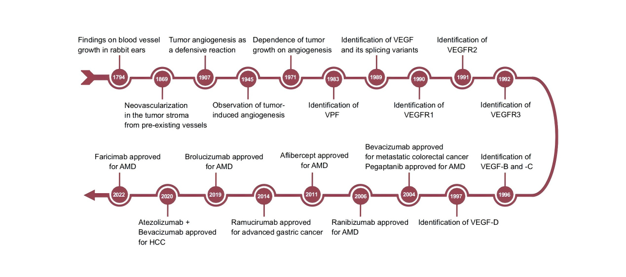
The discovery of VEGF (Vascular Endothelial Growth Factor) can be traced back over a century. In 1794, John Hunter, a British surgeon, first observed blood vessel growth in the ears of rabbits exposed to a cold environment [1]. Between 1939 and 1956, in the context of diabetic retinopathy, it was proposed that Diffuse Angiogenic Factors are involved in the formation of new blood vessels, and the specific term "Factor X" was coined to describe such molecules. Ide and colleagues were the first to suggest that a certain vascular growth-stimulating factor might provide nutrients for tumor growth [2]. A few years later, Algire and others found that tumors grew faster in areas with increased blood vessel density, and hypothesized that tumor growth might depend on an abundant blood supply [3].
In 1983, Senger's team extracted "Vascular Permeability Factor (VPF)" from the culture supernatant of guinea pig tumor cell lines, but the factor had not yet been isolated and sequenced. In 1989, the Ferrara Laboratory at Genentech reported the isolation and cloning of a heparin-binding endothelial cell mitogen protein from the culture medium of bovine pituitary follicular cells, thereby coining the technical term "VEGF" to describe this novel 45 kDa heparin-binding endothelial cell mitogen protein. In 1990, the heparin-binding endothelial cell mitogen protein was sequenced, VPF was purified and partially isolated, and it was matched with VEGF, confirming that VEGF and VPF are actually the same molecule.
In 1993, Kim found that neutralizing anti-VEGF antibodies could significantly reduce angiogenesis and the growth of tumor cells implanted in immunodeficient mice, which for the first time confirmed the therapeutic potential of targeting VEGF and opened up new therapeutic opportunities. In 1996, Carmeliet and others demonstrated that the inactivation of a single allele of the VEGF gene in mice led to vascular development defects and early embryonic death, highlighting the importance of VEGF in embryonic development [4]. In 2004, Bevacizumab was approved by the FDA for the treatment of metastatic colorectal cancer, becoming the first approved VEGF-targeted drug. Subsequently, Bevacizumab and other VEGF inhibitors (such as Aflibercept and Ranibizumab) were successively approved for the treatment of various cancers including non-small cell lung cancer, renal cancer, and glioblastoma, as well as diseases such as wet age-related macular degeneration (AMD) and diabetic retinopathy (DR). In 2018, the combination of Bevacizumab and a PD-L1 inhibitor (Atezolizumab) was approved for non-squamous non-small cell lung cancer, marking the entry of the synergistic application of VEGF and immunotherapy into clinical practice [5].
Overview of the VEGF Family
|
Family Member |
Main Functions |
Clinical Relevance |
|
VEGF-A |
Core factor regulating angiogenesis and vascular permeability |
Core target for anti-VEGF therapy (Bevacizumab, Ranibizumab) |
|
VEGF-B |
Metabolic regulation, myocardial protection |
Potential target for myocardial ischemia and diabetic cardiomyopathy |
|
VEGF-C |
Lymphangiogenesis, vascular remodeling |
Lymphedema, lymphatic metastasis of tumors |
|
VEGF-D |
Lymphangiogenesis, tumor metastasis |
Marker for lymphatic metastasis of breast cancer and lung cancer |
|
PlGF |
Placental development, inflammation, tumor angiogenesis |
Biomarker for preeclampsia (sFlt-1/PlGF ratio) |
|
VEGF-E (virus-derived) [6] |
Virus-induced angiogenesis |
Research model |

✅ Why Is VEGF-A Often Abbreviated as "VEGF"?
|
Dimension |
Explanation |
|
First discovered |
The "VEGF" first isolated from bovine pituitary glands by Ferrara and others in 1989 is actually VEGF-A165. |
|
Most core function |
VEGF-A is the major effector factor of "angiogenesis", regulating endothelial cell proliferation, migration, and permeability. Almost all physiological and pathological neovascularization depends on it. |
|
Most extensively studied |
Over 90% of VEGF-related research focuses on VEGF-A, and anti-VEGF therapies (such as Bevacizumab and Ranibizumab) all target VEGF-A. |
|
Most clinically relevant |
In diseases such as tumors, diabetic retinopathy, and AMD, VEGF-A is the main therapeutic target. |
✅ When Can Say "VEGF-A = VEGF"?
|
Scenario |
Whether They Can Be Equated |
|
"VEGF" as an abbreviation in literature/clinical reports |
✅Usually refers to VEGF-A |
|
Mechanism research, gene naming |
❌Need to clearly write VEGF-A |
|
Involving lymphatic vessels, metabolism, placental function |
❌Need to distinguish from other family members |
|
Description of anti-VEGF drug targets |
✅Default to targeting VEGF-A |
Whether in cell culture and protein detection in basic scientific research, or in pathological analysis and drug development in clinical translation, the "one-character difference" between VEGF and VEGF-A often determines the accuracy of experimental data and the reliability of research results. As the "general term" for the vascular endothelial growth factor family, VEGF actually includes multiple subtypes, while VEGF-A is the core member that regulates angiogenesis and participates in the occurrence of diseases——the misuse of antibodies in cell experiments or the confusion of indicators in clinical detection may lead to deviations in research directions or invalidation of data.
As an enterprise focusing on biological research tools and technical support, we are well aware of the importance of accurately distinguishing molecular targets for research. In the future, we will continue to provide more popular science on molecular biology knowledge and experimental technical guidelines to help researchers advance their research efficiently. If you encounter problems in VEGF/VEGF-A-related experiments or research, please feel free to leave a message for consultation, and we will provide you with professional answers!
References:
1.Lee C, Kim M J, Kumar A, et al. Vascular endothelial growth factor signaling in health and disease: from molecular mechanisms to therapeutic perspectives[J]. Signal Transduct Target Ther, 2025, 10: 170. https://doi.org/10.1038/s41392-025-02249-0
2.Ide A G, Baker N H, Warren S L. Vascularization of the Brown Pearce rabbit epithelioma transplant as seen in the transparent ear chamber[J]. Am J Roentgenol, 1939, 42: 891-899.
3.Algire G H, Chalkley H W, Legallais F Y, et al. Vascular reactions of normal and malignant tissues in vivo. I. Vascular reactions of mice to wounds and to normal and neoplastic transplants[J]. J Natl Cancer Inst, 1945, 6: 73-85.
4.Carmeliet P, Ferreira V, Breier G, et al. Abnormal blood vessel development and lethality in embryos lacking a single VEGF allele[J]. Nature, 1996, 380: 435-439.
5.Apte R S, Chen D S, Ferrara N. VEGF in Signaling and Disease: Beyond Discovery and Development[J]. Cell, 2019, 176(6): 1248-1264. https://doi.org/10.1016/j.cell.2019.01.021
6.Ogawa S, Oku A, Sawano A, et al. A Novel Type of Vascular Endothelial Growth Factor, VEGF-E (NZ-7 VEGF), Preferentially Utilizes KDR/Flk-1 Receptor and Carries a Potent Mitotic Activity without Heparin-binding Domain[J]. Journal of Biological Chemistry, 1998, 273(47): 31273-31282